


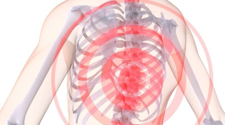
Osteochondrosis is a disease of the spine characterized by degenerative dystrophic damage to the intervertebral discs, vertebral bodies, and ligaments.
Spinal osteochondrosis has a chronic progressive course.The disease does not last long and symptoms only appear when complications arise.
According to the World Health Organization, 40-80% of the global population suffers from osteochondrosis.
The patients are mainly people over 30 years old.However, recently there has been a trend towards younger patients with osteochondrosis.Osteochondrosis ranks first among spinal diseases that disable patients.
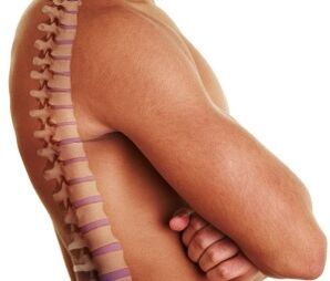
Brief anatomy of the spine
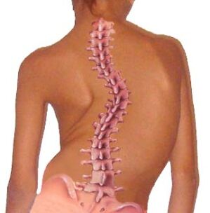
The spine performs the primary functions of spinal canal, support and movement, and also connects the head, shoulders and pelvic girdle.
The structural unit of the spine is the vertebrae.
The 24 vertebrae are connected to each other by intervertebral discs, which are the body's shock absorbers.
The spine is divided into five parts: cervical, thoracic, lumbar, sacral, and coccyx.
The normal spine shape is S-shaped.
This configuration of the organs distributes body weight and load evenly.
Structural and functional elements of the spine
Vertebrae are bony structures composed of vertebral bodies, vertebral arches, and processes.
The main load falls on the vertebral body, so this is its heaviest part.
Important!The arches of adjacent vertebrae form the spinal canal—the container for the spinal cord, blood vessels, spinal nerve roots, and fatty tissue.
ligamentThe spine is represented by the posterior longitudinal ligament, which connects the vertebrae along their posterior surface, and the ligamentum flavum, whose main purpose is to connect the arches of the vertebrae.
vertebrae.The vertebrae have seven processes extending from the vertebral arch: the spinous process, two transverse processes, two superior articular processes, and two inferior articular processes.The ligaments and muscles of the spine attach to the spinous processes.Other processes form the intervertebral joints of the spine.
intervertebral discIt is a disc-shaped plate composed of cartilage plate, annulus fibrosus and nucleus pulposus.Intervertebral discs connect adjacent vertebrae and provide mobility and stability to the spine.
intervertebral jointMade up of the processes of two adjacent vertebrae.The primary function of the intervertebral joints is to move the vertebrae relative to each other and provide flexibility to the spine.
intervertebral foramenLocated on the outside of the spine, it is formed by the articular processes, vertebral bodies, and pedicles of adjacent vertebrae.The spinal nerve roots exit through the intervertebral foramen and the blood vessels enter.
spinal cord- This is part of the central nervous system and consists of nerve fibers.The spinal cord has three membranes—the pia mater, the arachnoid mater, and the dura mater.The dura mater is made up of two sheets that connect and form the dural sac, which is filled with cerebrospinal fluid—cerebrospinal fluid.
spinal nerve root- These are the conductors of nerve impulses from the spinal cord to the internal organs and vice versa.Each spinal nerve root has autonomic, sensory, and nerve fibers within its structure.
paravertebral muscles- These are the muscles of the spine that support the spine and provide tilt and rotation of the body.
The functional unit of the spine isspinal motion segments, consisting of two adjacent vertebrae, discs, ligaments, and muscles.
Pathogenesis (developmental mechanism) of spinal osteochondrosis
Osteochondrosis occurs during developmentFour stages:
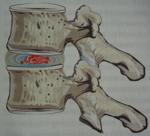
- Stage one.Pathological changes do not extend beyond the boundaries of the intervertebral disc.The nucleus pulposus dries out, causing the disc height to decrease.The annulus fibrosus cannot withstand the load - it ruptures and tears.
- The second stage.As the height of the intervertebral discs decreases, the spinal ligaments and muscles sag, resulting in instability of the spinal motion segments.Vertebrae can slide and move relative to each other.In this case, spondylolisthesis develops.
- The third stage.The condition is progressing.Intervertebral disc herniation and arthropathy of the intervertebral and uncinate joints occur.
- The fourth stage.During this phase, adaptive responses are activated in the form of vertebral bone growth (osteophytes).Therefore, the body attempts to limit excessive movement of the vertebrae.The sharp edges of osteophytes can damage spinal nerve roots.Fibrous ankylosis of the discs and joints develops, and the spine becomes immobile.The tonic phase is characterized by the disappearance of pain.
What causes osteochondrosis?
Osteochondrosis of the backIt is a multifactorial disease and it is impossible to pinpoint a specific cause.
The basis of osteochondrosis is a disruption of the microcirculation and metabolism of spinal tissues, which may be caused by improper load distribution on the spine.
Factors leading to osteochondrosis include:
- Improper posture in childhood (scoliosis, kyphosis, kyphosis, stooping);
- Back muscle weakness (spinal muscle weakness);
- Staying in one position for a long time (working in front of a computer, working in an office, doing manual work);
- Improper lifting;
- Lack of physical activity and sedentary lifestyle;
- Metabolic pathologies, especially calcium, phosphorus, calcium, vitamin, magnesium, zinc deficiencies;
- Genetic predisposition to osteochondrosis;
- infectious disease;
- The body is often hypothermic;
- chronic stress;
- hormonal imbalance;
- weightlifting;
- spinal injuries;
- Overweight and obesity.
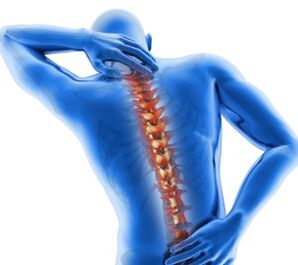
Symptoms of osteochondrosis
Chronic osteochondrosis can present with a variety of symptoms.It all depends on the stage of the disease, the extent of spinal damage, and the presence of complications.
Clinically, the disease appears when the degenerative dystrophic process has reached the posterior part of the annulus fibrosus and the posterior longitudinal ligament, and then the spinal nerve roots are irritated and compressed, and the conduction of nerve impulses is interfered with.
At the same time, the spinal cord and blood vessels will also be compressed, manifesting as reflex compression syndrome.
Important!The pain syndrome in osteochondrosis occurs because osteophytes, spastic muscles, and displaced vertebrae compress the spinal nerve roots in the intervertebral foramina.
Osteochondrosis and its symptoms often mimic acute coronary syndrome, pleurisy, acute pancreatitis, hepatic and renal colic, acute appendicitis, and adnexitis.
Therefore, it is important to perform a thorough differential diagnosis of the disease to rule out life-threatening conditions.
most commonSymptoms of osteochondrosis:
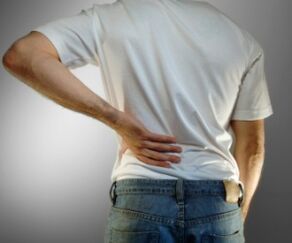
- Neck, waist, and thoracic spine pain, which may be aching, throbbing, or lower back pain.The pain radiates to the head, upper limbs, lower limbs, shoulder blades, heart, and stomach.Pain syndrome may worsen with physical activity, sneezing, laughing, coughing, or remaining in one position for long periods of time;
- sensory impairmentDifferent parts of the body are at the level of innervation of the pinched nerve;
- Crampmuscles of the neck, back, upper and lower limbs;
- Migraine-like Headache;
- painat the joints of the limbs;
- increased fatiguefrom physical and mental labor;
- Dizziness and loss of consciousnessRapid head rotation (vertebral artery syndrome);
- visual impairment(floaters or colored spots in front of the eyes);
- Hearing loss, tinnitus;
- Heartache;
- painalong the intercostal spaces;
- reduced blood supplyUpper and lower limbs, manifested by cold skin;
- Paresthesia– Crawling, tingling and burning sensations in the spine;
- dry skin;
- sweating disorder;
- urination disorder(difficulty urinating, enuresis);
- Loss of libido, impotence.
Early diagnosis of osteochondrosis will greatly facilitate its treatment.
How to Diagnose Osteochondrosis
Neuropathologists diagnose osteochondrosis.If necessary, patients can be referred to cardiologists, gastroenterologists, orthopedists, surgeons, etc. for consultation.
During the interview, it is necessary to determine exactly the nature of the complaint, when it occurred and what the patient associates it with.Be sure to check the medical history, the patient's occupation, and whether close relatives have osteochondrosis.
In this case, laboratory testing provides no information.By conducting blood biochemistry tests, you can pay attention to the levels of trace elements such as calcium and phosphorus.
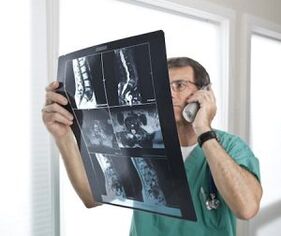
The main methods in the diagnosis of osteochondrosis are instrumental methods such as spinal radiography, computed tomography, and magnetic resonance imaging.
X-rays of the spine are the easiest, most convenient, and informative way to diagnose osteochondrosis.
Mandatory radiography is a direct and lateral projection of the desired portion of the spine.Osteochondrosis is characterized by reduced disc height, the presence of osteophytes, osteoporosis, and spinal deformity.
myelography- This is an X-ray examination of the spine by introducing a contrast agent into the spinal canal.This method is dangerous because of allergic reactions to the contrast medium.
Myelography allows us to study the internal structures of the spinal canal.This method is valuable in diagnosing Schmohl's hernia (intervertebral hernia).
Computerized and MRI scans– These are modern diagnostic methods that allow layer-by-layer visualization of the soft tissue and bones of the spine.
These methods are expensive and are therefore used in the differential diagnosis of severe cases, especially osteochondrosis and diseases with similar symptoms.
Because osteochondrosis often masquerades as diseases of the heart, lungs, pleura, stomach, intestines, kidneys, and liver, differential diagnosis is required.
For this purpose, the patient may be subjected to an electrocardiogram, an ultrasound of the heart and internal organs, a troponin blood test, a vascular ultrasound, a chest X-ray, an electroencephalogram, etc.
Treatment methods for osteochondrosis
Osteochondrosis treatment canConservative and surgical.
Important!First, a comprehensive conservative approach is adopted, with surgical treatment being resorted to only in extreme cases.
Let's consider how to properly treat osteochondrosis.KconservativeTreatment methods for osteochondrosis include:
- drug treatment;
- physical therapy;
- physical therapy methods;
- manual therapy;
- massage;
- acupuncture.
drug treatmentThe goals of osteochondral therapy are to relieve pain, relax muscles, relieve nerve and muscle swelling, and improve blood flow and conduction of nerve impulses.For this purpose, the following groups of drugs are used:
- NSAIDs;
- chondroprotectant, which includes components of cartilage tissue.These medications protect the cartilage of the vertebrae and discs from the negative effects of a variety of factors;
- diureticsRemoves excess fluid from the body and relieves swelling of spinal nerve roots and paravertebral muscles;
- muscle relaxantsRelax cramped muscles;
- drug, improve the metabolism and microcirculation of spinal tissue (vitamins B1, B6, B12, C, A and E);
- Calcium supplements;
- Hormone drugs, prescribed when NSAIDs are ineffective.
therapeutic exercise– These are quantitative physical activities that can be performed at home and at work to treat and prevent osteochondrosis.
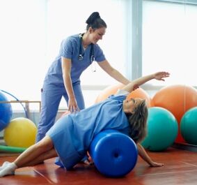
There are many sets of exercises for osteochondrosis.The prescription of exercise therapy and the monitoring of its implementation are carried out by a qualified specialist - a physiotherapist.
With correctly chosen exercise therapy, you can relieve pain, improve mobility and blood supply to the spine, and halt the progression of the disease.
physical therapyOsteochondral treatment is performed by physiotherapists in special physiotherapy departments of hospitals, nursing homes and pharmacies.
Physical therapy methods include: electrophoresis, magnet therapy, laser therapy, mud therapy, bath therapy, ultraviolet irradiation of the affected part of the spine, vibration therapy, etc.
manual therapy– This is a measured manual impact on the spine to restore its mobility, eliminating displacement of vertebrae and discs.
Manual therapy should only be performed by a qualified chiropractor.
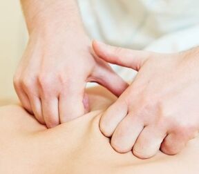
Massage and self-massageFor osteochondrosis, its purpose is to relieve muscle spasms, improve microcirculation in paravertebral tissues, and increase spinal mobility.
AcupunctureIt is a method of treating osteochondrosis by injecting fine needles into active points.
Under the action of acupuncture, the levels of endogenous opioids and cortisol in the body increase, which have anti-inflammatory and analgesic effects.
Prevent osteochondrosis
To maintain your health and keep your spine mobile into old age, follow these principles for preventing osteochondrosis:
- pay attention to your posture– Always keep your back straight and don’t hunch;
- choosecorrect posturefor sleeping;
- Sit correctly at the table(Shoulders should be relaxed, back straight, and furniture should fit your height);
- When holding one position for an extended period of time (in the office, at the computer, sitting in front of a craft), try every 1-1.5 hoursdo some physical exercise, self-massage your back, or just get up and take a walk;
- Distribute the load correctlythe impact on the spine when lifting and carrying various heavy objects;
- wear orthopedic shoes;
- healthy sleepPlace on a flat, firm to medium-firm mattress.It is best to buy orthopedic mattresses and pillows.
spinal osteochondrosisUnfortunately, this is a chronic progressive disease and there is no cure.The effectiveness of treatment directly depends on its timeliness.
Do not self-medicate to avoid worsening of the condition.At the first sign of osteochondrosis, contact a neurologist.